Microscopy Suite
Location: Chaparral Hall, Room 5434; (818) 677-4575.
For information, training, and reservations, contact Dr. Ernest Kwok: 818-677-3383; ernest.kwok@csun.edu.
Click HERE to access calendar for using confocal microscope. Click HERE to access pre-training eLessons that you should master before scheduling hands-on training.
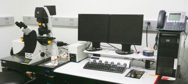
Confocal Microscope
Leica TCS SP5 II confocal microscope, good for taking images of living cells that have components marked with fluorophores. Lasers allow for optical sectioning and construction of 3D images. This microscope is equipped with 4 lasers providing a total of 7 excitation wavelengths: 405, 458, 476, 488, 514, 543, and 633 nm. Confocal fluorescence detection is by 3 independently adjustable PMTs. This microscope is also equipped with 4 epifluorescence filter sets (images cannot be collected at this time).
| filter set/fluorophore | Excitation | Dichronic | Emission |
| DAPI/Hoechst | 340-380 nm | 400 | >425 |
| GFP/FITC | 450-490 | 495 | 500-550 |
| GFP/FITC LP | 450-490 | 510 | >515 |
| Rhodamine/TRITC | 515-560 | 580 | >590 |
For Transmitted Light microscopy, the microscope is capable of Brightfield and Differential Interference Contrast (DIC) using a dedicated PMT.
Brightfield & Phase Contrast
 Zeiss AxioObserver Z1 inverted epifluorescence microscope. Location Citrus Hall, Room 3219.
| Filter Set/fluorophore | Excitation | Dichroic | Emission |
| DAPI/Hoechst | 300-400 nm | 395 | 420-470 |
| GFP/FITC | 450-490 | 495 | 500-550 |
| YFP | 488-513 | 515 | 520-550 |
| Rhodamine/TRITC | 533-558 | 570 | 570-640 |
For Transmitted Light microscopy, the microscope is capable of Brightfield, Phase Contrast (10x objective) and Differential Interference Contrast (DIC; 40x and 63x objectives).
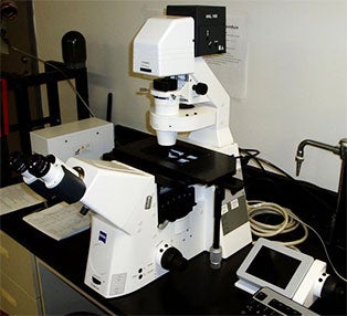
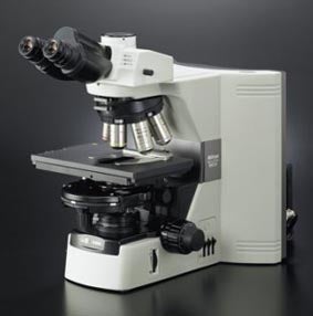
Fluorescent Microscopes
Left: fluorescent microscope with 2 filter sets.
| Filter Set/fluorophore | Excitation | Dichroic | Emission |
| GFP/FITC | 450-490 nm | 495 | 500-550 |
| Rhadamine/TRITC | 510-550 | 570 | >560 |
Right: Nikon 80-i microscope equipped with a Jenoptik 14+ 12MP camera, phase contrast, brightfield, polarized light and DIC for transmitted light observation.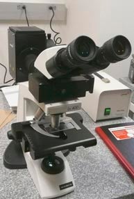
Ultramicrotome
Ultramicrotome makes very, very thin sections to be studied with transmission electron microscope.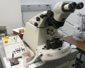
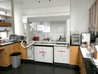
Fume Hood
Fume hood area of microscopy suite.