Garrett Research Group: Experimental Techniques
Images
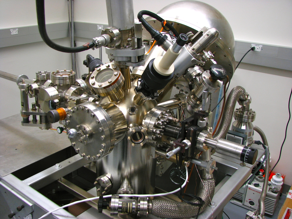
X-ray Photoelectron Spectroscopy (XPS)
In XPS, we irradiate a solid surface with soft X-rays and measure the kinetic energy of the electrons emerging from the surface. Since each atom has a unique set of atomic orbitals, the energy of the photoelectrons is diagnostic of that element. The technique is quantitative and measures atomic composition of all elements (except H and He) within the uppermost few atomic layers. Small shifts in the kinetic energy of the departing photoelectrons can indicate the oxidation state of the element, for example distinguishing metallic titanium from its ionic compounds. The technique is conducted in a vacuum chamber and the instrument (Physical Electronics PHI 5200) is located in the Garrett research laboratory.
Images
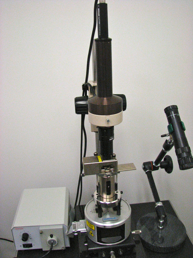
Atomic Force Microscopy (AFM)
Our atomic force microscope (Bruker MultiMode V) moves a sharp tip attached to a flexible cantilever across a surface to measure its topology. The tip can be made to contact the surface, hover closely above it or to vibrate, "tapping" the surface as it moves. In all cases, displacement of the tip/cantilever provides an image of the surface in three dimensions. In optimal conditions, the technique is capable of imaging individual atoms although in our laboratory the features are usually from a few micrometers to a few tens of nanometers in size. The instrument is sensitive to acoustic and mechanical vibrations so is mounted on an air table and in a small separate room in the Garrett research laboratory.
Images
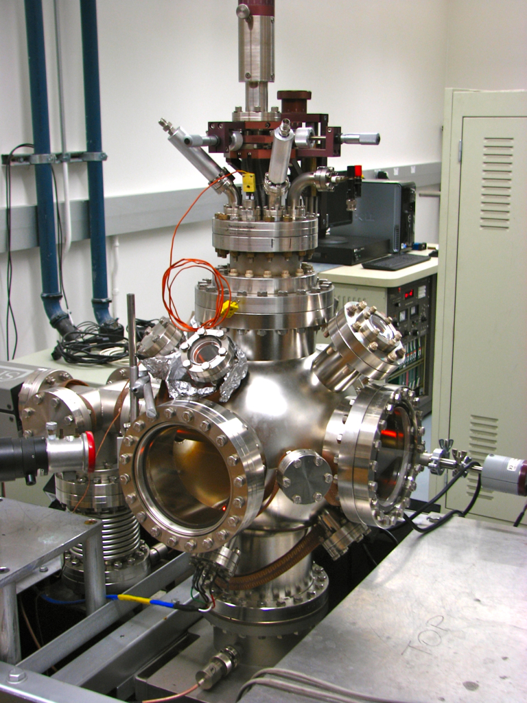
Mass Spectrometry
An electron-impact mass spectrometer fragments a gas phase molecule to produce a slew of fragments with masses abundances characteristic of the initial molecule. We can use this information to identify unknown molecules, usually by comparison with a standard mass spectrum. We use a mass spectrometer (Stanford Research Systems RGA 200) in conjunction with a coolible/heatable surface. Molecules are condensed onto a cold surface in vacuum. After some treatment, the surface is heated until the molecules desorb and are caught by the mass spectrometer, a technique known as temperature programmed desorption (TPD). Thermodynamic and kinetic information can be obtained in addition to the products of reactions. This instrument was constructed in-house and is located in the Garrett research laboratory.
Images

Electrochemistry
Corrosion studies amenable to investigation by electrochemistry. In one approach, termed voltammetry (potentiostatic and potentiodynamic polarization), we apply a potential to the material under investigation in a corrosive medium and monitor the current that flows due to oxidation or reduction reactions. In general, the more current that flows, the more rapid the corrosion. When no external potential is applied, information on the intrinsic corrosion rate and thermodynamic stability of the material are obtained. Such information is crucial when designing corrosion-resistant alloys. An electrochemical potentiostat (EG&G 273A) and three-electrode cell system is located in the Garrett research laboratory.
Images
Other Techniques
Rather than base our research on one or two kinds of instruments, we focus on chemical problems and find the best instruments to answer questions. As such, students in the group use many different instruments, not only those based in the Garrett research laboratory. These include X-ray diffraction (XRD), scanning electron microscopy with energy-dispersive spectroscopy (SEM-EDS), transmission electron microscopy (TEM), inductively-coupled mass plasma spectrometry (ICP-MS), ultraviolet-visible spectroscopy (UV-Vis) and differential scanning calorimetry (DSC) as the need arises.
Last updated 11/24/19.