I. INTRODUCTION
In this lesson, you will investigate some properties of skeletal muscle. Physiological phenomena associated with other kinds of muscle, such as electrophysiology of the heart, will be studied in subsequent lessons.
The human body contains three kinds of muscle tissue and
each performs specific tasks to maintain homeostasis:, Cardiac muscle,
Smooth muscle, and Skeletal muscle.
Cardiac muscle is found only in the heart. When it contracts, blood circulates, delivering nutrients to cells and removing cell waste.
Smooth muscle is located in the walls of hollow organs, such as the intestines, blood vessels or lungs. Contraction of smooth muscle changes the internal diameter of hollow organs, and is thereby used to regulate the passage of material through the digestive tract, control blood pressure and flow, or regulate airflow during the respiratory cycle.Skeletal muscle derives its name from the fact that it is usually attached to the skeleton. Contraction of skeletal muscle moves one part of the body with respect to another part, as in flexing the forearm. Contraction of several skeletal muscles in a coordinated manner moves the entire body in its environment, as in walking or swimming.
The primary function of muscle, regardless of the
kind, is to convert chemical energy to mechanical work, and in so
doing, the muscle shortens or contracts.
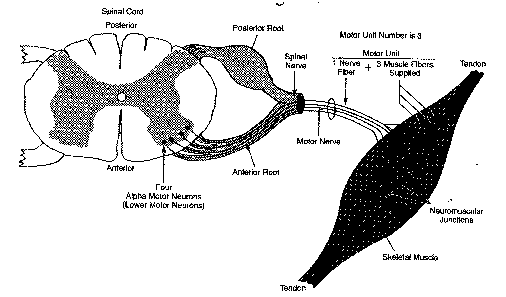
Human skeletal muscle consists of hundreds of individual cylindrically shaped cells (called fibers) bound together by connective tissue. In the body, skeletal muscles are stimulated to contract by somatic motor nerves that carry signals in the form of nerve impulses from the brain or spinal cord to the skeletal muscles (Fig. 1.1). Axons (or nerve fibers) are long cylindrical extensions of the neurons. Axons leave the spinal cord via spinal nerves and the brain via cranial nerves, and are distributed to appropriate skeletal muscles in the form of a peripheral nerve, which is a cable-like collection of individual nerve fibers. Upon reaching the muscle, each nerve fiber branches and innervates several individual muscle fibers.

The size of the motor unit arrangement of a skeletal muscle (e.g., 1:10, 1:50, or 1:3000) is determined by its function (flexion, extension, etc.) and location in the body. The smaller the size of a muscle's motor units, the greater the number of neurons needed for control of the muscle, and the greater the degree of control the brain has over the extent of shortening. For example, muscles which move the fingers have very small motor units to allow for precise control, as when operating a computer keyboard. Muscles that maintain posture of the spine have very large motor units, since precise control over the extent of shortening is not necessary.
Physiologically, the degree of skeletal muscle contraction is controlled by:
1. Activating a desired number of motor units within the muscle, and
2. Controlling the frequency of motor neuron impulses in each motor unit.
When an increase in the strength of a muscle's contraction is necessary to perform a task, the brain increases the number of simultaneously active motor units within the muscle. This process is known as motor unit recruitment.
Resting skeletal muscles in vivo exhibit a phenomenon known as tonus, a constant state of slight tension that serves to maintain the muscle in a state of readiness. Tonus is due to alternate periodic activation of a small number of motor units within the muscle by motor centers in the brain and spinal cord. Smooth controlled movements of the body (such as walking, swimming or jogging) are produced by graded contractions of skeletal muscle. Grading means changing the strength of muscle contraction or the extent of shortening in proportion to the load placed on the muscle. Skeletal muscles are thus able to react to different loads accordingly. For example, the effort of muscles used in walking on level ground is less than the effort those same muscles expend in climbing stairs.
When a motor unit is activated, the component muscle fibers generate and conduct their own electrical impulses that ultimately result in contraction of the fibers. Although the electrical impulse generated and conducted by each fiber is very weak (less than 100 microvolts), many fibers conducting simultaneously induce voltage differences in the overlying skin that are large enough to be detected by a pair of surface electrodes. The detection, amplification, and recording of changes in skin voltage produced by underlying skeletal muscle contraction is called electromyography. The recording thus obtained is called an electromyogram (EMG).
II. EXPERIMENTAL OBJECTIVES
1) To observe and record skeletal muscle tonus as reflected by a basal level of electrical activity associated with the muscle in a resting state.
2) To record maximum clench strength for right and left hands.
3) To observe, record, and correlate motor unit recruitment with increased power of skeletal muscle contraction.
4) To listen to EMG "sounds" and correlate sound intensity
with motor unit recruitment.