I. INTRODUCTION
The brain constantly receives sensory input and integrates
the information. The sensory information is relayed from the periphery
through lower centers in the brain, and then the information is sent to
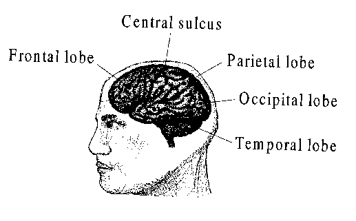
specific regions of the cerebral cortex where it is processed. For
example, the occipital lobe processes visual information while the
parietal
lobe processes non-visual, sensory information such as cutaneous pain
(Fig 4.1). If you choose to, you can direct your attention to particular
bits of sensory information; you can access memories associated with the
sensory information; or you can selectively ignore this sensory input.
The blood/brain barrier separates cerebral spinal fluid from the blood. Oxygen, glucose, and carbon dioxide can cross the blood/brain barrier, but the hydrogen ion can not. The brain requires oxygen and glucose for energy. Without a relatively constant source of oxygen and glucose, the brain ceases to function. Levels of carbon dioxide in the spinal fluid can change the pH of the spinal fluid, which can in turn change the body's respiration rate.
Because brain activity is related to ions and charge movement, this activity can be detected by electrodes. The record of the brain's activity is called an electroencephalogram (EEG) from the root words of electro (electrical), encephalo (brain), and gram (record).
The EEG records the electrical activity on the surface
of the cerebral cortex. The EEG is complex and variable between adults,
although under certain conditions, the EEG exhibits simpler, rhythmic activity.
Simpler patterns in the EEG occur when many cells synchronize their
input to the surface of the cerebral cortex. The more synchronized the
charge movement, the more rhythmic the EEG.
Your EEG changes as you grow. The development of EEG is rapid with newborns. As neural development proceeds, the EEG recorded from the posterior regions of the brain of an infant of 3-4 months begins to resemble EEGs recorded from the posterior region of adults. The difference is that the 3-4 month old infants have EEGs in the frequency range of 3-4 Hz, whereas adults tend to have average frequencies of 10 Hz. By the time the infant is one year old, the posterior region EEG is approximately 6 Hz, by three years, 8 Hz, and by 13-14 years (puberty), the average frequency is 10 Hz (similar to adults).
One of the simpler patterns is the alpha rhythm.
The alpha rhythm is characterized by a frequency of 8-13 Hz and amplitudes
of 20-200 uV. Each region of the brain has a characteristic frequency of
alpha rhythm. Alpha waves of the greatest amplitude tend to be recorded
from the occipital and parietal regions of the cerebral cortex.
Just as the EEG is variable depending on the mental state of an individual, the frequency and amplitude of alpha rhythms within an individual change. In general, the alpha rhythm is the prominent EEG wave pattern of an adult in a relaxed, inattentive state with eyes closed.
More specific conditions of alpha rhythms are listed below:
In this lesson, you will record the EEG and alpha
rhythm under several conditions. At the same time, the root-mean-squared
of the alpha rhythm (alpha-RMS) and an "alpha thermometer" will
be displayed. Alpha-RMS and the "alpha thermometer" are indices of the
activity levels of the alpha rhythm.