Lab 2
Back to Table
of Contents
SHEEP BRAIN DISSECTION: LAB 2
External Features:
There are several systems for subdividing the brain.
The outline presented in TABLE
2 provides a very useful format for
studying the brain as a set of divisions that emerged during evolution. The
CNS is arranged in a stratified, or layered manner. The strata were added
as the nervous system evolved from a primitive neural tube to the elaborate
structure we know as the human brain. The lower, or caudal, strata,
generally, are involved with less complex neural activities than the higher,
or more rostral, strata above them.
Ventricular System:
When in the skull, the external surface of the brain
is awash in a bath of CSF. There are also a number of CSF-filled cavities
that are located on both the external surface of the brain and in its interior.
The fluid-filled cavities located on the external surface of the brain are called
cisterns, while
the fluid-filled cavities located internally are called ventricles.
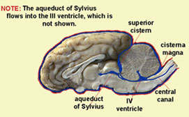 When the dura mater was in place, it formed an enclosed space
at the most ventro-caudal point of the cerebellum, just above its junction with
the medulla and the pons.
This space is called the cisterna magna.
In the intact brain it is filled with cerebrospinal fluid. The meninges
form another enclosed space at the anterior limit of the cerebellum called the
superior cistern. Use the links
to the images that show where these two fluid filled chambers were located in
the intact brain. (Click on the image for an enlarged view.)
When the dura mater was in place, it formed an enclosed space
at the most ventro-caudal point of the cerebellum, just above its junction with
the medulla and the pons.
This space is called the cisterna magna.
In the intact brain it is filled with cerebrospinal fluid. The meninges
form another enclosed space at the anterior limit of the cerebellum called the
superior cistern. Use the links
to the images that show where these two fluid filled chambers were located in
the intact brain. (Click on the image for an enlarged view.)
As discussed in the lecture and your textbook, the brain evolved from
a primitive neural tube. As certain segments of the tube enlarged, the internal
spaces in the tube followed suit. This resulted in the creation of large
spaces in the interior of the brain called ventricles.
The ventricles and their connecting passages are filled with the same CSF as
that in the subarachnoid space. The ventricles are connected to one another
and to the subarachnoid space by apertures (openings or windows) called foramina
(singular = foramen). Thus, the brain is
cushioned by CSF that fills the ventricles within and the cisterns and subarachnoid
space without.
At the lateral junction of the cerebellum and the medulla notice the dark-brown,
tufted material, the choroid plexus.
This material is a capillary bed, which, along with other tissue, is involved
in the production of CSF. We will see another choroid plexus in the lateral
ventricles in Lab 3.
Important structures or features of the dorsal
surface of the brain.
 The
dominant features of your sheep brain specimen are the two cerebral
hemispheres and the exquisitely convoluted
cerebellum. The
cerebral hemispheres are divided into functional subsections called lobes
or poles. To see the four lobes of neocortex of the sheep identified,
follow the link. The lobes of the brain are separated from one another by sulci,
or fissures. Two important sulci can be found in the anterior regions
of the two hemispheres. Together, these two sulci form a 'T.' The stem
of the 'T' is made by the coronal sulcus, which runs parallel and
The
dominant features of your sheep brain specimen are the two cerebral
hemispheres and the exquisitely convoluted
cerebellum. The
cerebral hemispheres are divided into functional subsections called lobes
or poles. To see the four lobes of neocortex of the sheep identified,
follow the link. The lobes of the brain are separated from one another by sulci,
or fissures. Two important sulci can be found in the anterior regions
of the two hemispheres. Together, these two sulci form a 'T.' The stem
of the 'T' is made by the coronal sulcus, which runs parallel and 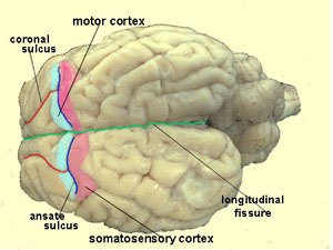 lateral
to the longitudinal fissure, the fissure that separates the two hemispheres.
(Note: the coronal sulcus seems to be ill-named, because it runs perpendicular
to the coronal plane of dissection. Don't be confused by this.)
The coronal sulcus divides the frontal poles into approximately equal
left- and right-halves. Follow the coronal sulcus caudally
until it ends at the ansate sulcus, which forms the head of the 'T.' The
frontal lobe is separated from the parietal lobe by the ansate
sulcus (called the central or Rolandic fissure in man). The
area anterior to the ansate sulcus is frontal lobe and the cortex posterior
to the ansate sulcus is the parietal lobe. The gyrus immediately anterior
to the ansate sulcus is the precentral gyrus.
The gyrus immediately posterior to the ansate sulcus is known as the postcentral
gyrus in the human brain. Classically, the precentral
gyrus is thought of as motor cortex, and
the postcentral gyrus as somatosensory cortex. It is more
correct to think of the two as being predominantly motor and predominantly somatosensory,
respectively, because other functions are also present.
lateral
to the longitudinal fissure, the fissure that separates the two hemispheres.
(Note: the coronal sulcus seems to be ill-named, because it runs perpendicular
to the coronal plane of dissection. Don't be confused by this.)
The coronal sulcus divides the frontal poles into approximately equal
left- and right-halves. Follow the coronal sulcus caudally
until it ends at the ansate sulcus, which forms the head of the 'T.' The
frontal lobe is separated from the parietal lobe by the ansate
sulcus (called the central or Rolandic fissure in man). The
area anterior to the ansate sulcus is frontal lobe and the cortex posterior
to the ansate sulcus is the parietal lobe. The gyrus immediately anterior
to the ansate sulcus is the precentral gyrus.
The gyrus immediately posterior to the ansate sulcus is known as the postcentral
gyrus in the human brain. Classically, the precentral
gyrus is thought of as motor cortex, and
the postcentral gyrus as somatosensory cortex. It is more
correct to think of the two as being predominantly motor and predominantly somatosensory,
respectively, because other functions are also present.
The separation
of the parietal lobe from the more posteriorly located occipital
lobe is ill-defined. The temporal lobe in the sheep is very
poorly developed in comparison to primates. The temporal lobe (sometimes
called insula in the sheep) is quite small as can be seen in the figure.
Before you continue, stop and look at the mounted
phylogenetic scale of brains and the preserved brains in jars that are available.
Notice that the single-most distinctive feature of the
phylogenetic sequence is the increase in the relative size of the cerebrum (cerebral
hemispheres). Notice that the frog's brain is distinguished from the codfish
by a noticeable increase in cerebrum. Compare the brain of the dog with that
of the cat and rat. Now, look at the human brain specimen.
Look at the dorsal aspect of the brain (and the spinal cord
that extends from it). The dorso-medial
sulcus marks the midline of the spinal cord. (Have the instructor
or lab assistant point this out to you.) You should be able to see that the
dorsal surface of the medulla, and 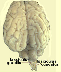 the
spinal cord, are marked by parallel, longitudinal striations formed by columns
of fibers. The most medial pair of these columns is the fasciculus
gracilis; the more lateral pair of columns is the fasciculus
cuneatus. (Have the instructor or lab assistant point these
out to you, if you are unclear about their locations.) Fasciculus means
tract. Collectively, these two fasciculi are also known as the dorsal
columns. The axons in these columns are ascending sensory fibers,
carrying for the most part, light touch sensations from the body and limbs.
The fasciculus gracilis conducts ipsilateral
information from the lower body and hind limbs, while the fasciculus
cuneatus conducts ipsilateral information from the upper body and
forelimbs. (Click on the image at the left for an expanded view.)
the
spinal cord, are marked by parallel, longitudinal striations formed by columns
of fibers. The most medial pair of these columns is the fasciculus
gracilis; the more lateral pair of columns is the fasciculus
cuneatus. (Have the instructor or lab assistant point these
out to you, if you are unclear about their locations.) Fasciculus means
tract. Collectively, these two fasciculi are also known as the dorsal
columns. The axons in these columns are ascending sensory fibers,
carrying for the most part, light touch sensations from the body and limbs.
The fasciculus gracilis conducts ipsilateral
information from the lower body and hind limbs, while the fasciculus
cuneatus conducts ipsilateral information from the upper body and
forelimbs. (Click on the image at the left for an expanded view.)
Follow the dorsal columns rostrally until they just begin to disappear beneath
the cerebellum; you will find two small mounds. These two swellings are
the nucleus gracilis and the
nucleus cuneatus. (Remember from Lab 1 that nuclei are collections
or clusters of cell bodies located within the CNS.) The axons in the dorsal
columns synapse on cells located in the nucleus gracilis and nucleus cuneatus.
The axons of the cells residing in the two nuclei then exit the nucleus, decussate
(cross the midline) and project to the contralateral thalamus where they synapse
on cells in the ventroposterolateral nucleus (VPL).
The axons of the cells located in VPL project to somatosensory
cortex (the post-central gyrus).
Before you continue, stop to draw a schematic of
the path that somatosensory information takes from spinal cord to postcentral
gyrus. Include the nucleus gracilis, nucleus cuneatus, VPL and postcentral
gyrus in your drawing.
Important features of the ventral
surface.
In Lab 1, you located a large fiber structure, the cerebral
peduncles, just anterior to the pons. The oculomotor nerves can
be seen exiting from them. Recall that the cell bodies that give rise to the
axons in the cerebral peduncles are found in motor cortex and that they are
called pyramidal cells. The fibers in the cerebral peduncles continue
to the spinal cord. They can be seen on the ventral surface of the medulla,
where they are known as the pyramidal
tract. The axons continue to spinal
cord where they split into three bundles; two of them are the lateral
corticospinal tract and the ventral
(or anterior) corticospinal
tract.
Stop, now, and mentally trace the pyramidal tract
from cerebral cortex to spinal cord.
The pyramidal motor system,
one of two major motor systems in the body is in control of fine, discrete and
voluntary motor activities such as writing, typing, or playing the piano. Other
motor systems are concerned with gross motor movements, such as, dancing, walking,
or waving goodbye.
This may be a good time to restate conventions concerning
names of tracts in the CNS. If you keep the rule 'from-to' in mind, you
will always be able to tell the site of origin and destination for a given tract.
The first name in the title indicates the site of origin of the tract, while
the second name indicates the tract's destination. The tract known as
the corticospinal tract, according to the rule, originates from neurons whose
cell bodies reside in the cortex and project their axons to the spinal cord.
Conversely, a tract called the spinothalamic tract originates from neurons in
the spinal cord and ends, or synapses, in the thalamus.
At the anterior end of the ventral surface of the medulla, immediately
caudal to the pons, locate a band of transverse fibers called the trapezoid
body. The trapezoid body consists
of fibers carrying information from the right ear to left auditory cortex and
information from the left ear to right auditory cortex. The trapezoid body is
to the auditory system what the optic chiasm is to the visual system.
Unlike somatosensory cortex, the auditory and visual cortices receive bilateral
input, that is, each projection site receives information from both ears or
both eyes, respectively. (If it has not been stripped away, you will find
the VIII cranial nerve, the vestibulo-cochlear or auditory nerve at the most
lateral extent of the trapezoid body.
On the ventral surface of your sheep brain, locate the
very prominent swelling between the trapezoid body and the cerebral peduncles,
the pons.
Its name is derived from the Latin word, pons, which means 'bridge.'
The structure is aptly named, because of the great number of decussating fibers
that cross the midline here and project to the cerebellum. In addition to the
decussating fibers in the pons, there are "fibers of passage," that is,
fibers that merely pass through the pons on their way to some other target without
synapsing in the pons. Pyramidal tract fibers descending from motor cortex
to their destination in the spinal cord are one example of these fibers of passage.
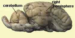
Immediately posterior to the the cerebral hemispheres, you find the cerebellum,
a large, complex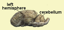 structure concerned with all levels of motor coordination. The cerebellum, which
means 'little brain,' also has an outer layer of cortex, that is,
it is covered by a multilayered mantle of cells like the layer of cortex on
the cerebral hemispheres. The cerebellar surface is characterized by intricate,
extremely fine convolutions called folia.
The folia are analogous to the gyri of the cerebral hemispheres. Like
the cerebral hemispheres, the cerebellum has an inner core of white matter.
In the cerebellum this inner core of white matter is called the arbor
vitae. (Click on the image below for a larger view.) The white-matter
core consists of axons projecting to and from the cerebellar hemispheres, the
spinal cord, sensory and motor cortices, and other regions of the brain.
The cerebellum sends information to the
structure concerned with all levels of motor coordination. The cerebellum, which
means 'little brain,' also has an outer layer of cortex, that is,
it is covered by a multilayered mantle of cells like the layer of cortex on
the cerebral hemispheres. The cerebellar surface is characterized by intricate,
extremely fine convolutions called folia.
The folia are analogous to the gyri of the cerebral hemispheres. Like
the cerebral hemispheres, the cerebellum has an inner core of white matter.
In the cerebellum this inner core of white matter is called the arbor
vitae. (Click on the image below for a larger view.) The white-matter
core consists of axons projecting to and from the cerebellar hemispheres, the
spinal cord, sensory and motor cortices, and other regions of the brain.
The cerebellum sends information to the 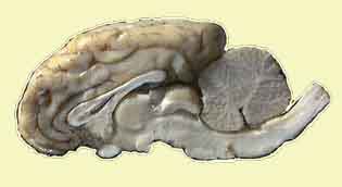 brain
and spinal cord via axons that exit from the cerebellum. In this way,
we have information coming into the cerebellum that helps guide cerebellar control
of our motor behavior. Without an intact cerebellum, you would find it
difficult to walk, maintain a sense of balance, or to perform a complex behavior,
such as, hit a tennis ball with a tennis racket, an action that requires hand-eye
coordination and timing. The cerebellum has other important functions; it is
important for establishing skill memories and for the occurrence of classically
conditioned responses.
brain
and spinal cord via axons that exit from the cerebellum. In this way,
we have information coming into the cerebellum that helps guide cerebellar control
of our motor behavior. Without an intact cerebellum, you would find it
difficult to walk, maintain a sense of balance, or to perform a complex behavior,
such as, hit a tennis ball with a tennis racket, an action that requires hand-eye
coordination and timing. The cerebellum has other important functions; it is
important for establishing skill memories and for the occurrence of classically
conditioned responses.
Nestled between the cerebellum and the cerebral hemispheres
are two prominent elevations sitting symmetrically on either side of the midline.
You may have to pull your cerebellum gently and caudally to reveal them. Collectively,
these four structures are called the corpora quadrigemina
('bodies of four twins'), but it is easier to remember them in their pairwise
configurations: the caudal and smaller pair, the inferior
colliculi, are part of the auditory system, while the larger, anterior
pair, the superior colliculi,
are part of the visual system. This region of the midbrain is also called
the tectum ('roof'), because the colliculi
('little hills') form the roof, or upper boundary of the Aqueduct of Sylvius. Look
at the figure connected to this link
to see the relationship between the colliculi (tectum) and the aqueduct of sylvius.
The figure will clarify the location of the colliculi. If you are unable to
see any of the structures named in this paragraph clearly and easily, seek help
from the instructor, or lab assistant.
On the ventral surface of the brain, at the midline just anterior
to the oculomotor nerve, locate the small, but distinct, tissue that looks like
the tip of a tongue. These are the mammillary
bodies, which mark the caudal limit of the hypothalamus.
Cells in the mammillary bodies are particularly vulnerable to alcohol.
Autopsies have shown significant destruction of the mammillary bodies in chronic
alcoholics suffering from a severe memory disorder known as Korsakoff's syndrome.
Some neurologists believe that the mammillary bodies are involved in memory
processes.
While the mammillary bodies form the caudal limit of the hypothalamus,
its anterior border is marked by the optic chiasm. The lateral boundaries
of the hypothalamus are rimmed by the medial edges of the cerebral peduncles.
The general outline of the hypothalamus from the ventral aspect, thus, assumes
a diamond-like configuration. Although the hypothalamus is not a very
large structure, it is quite complex. The hypothalamus contains many different
nuclei that are concerned with regulation of temperature, hunger and satiety,
sexual behavior, and, perhaps, even sexual preference.
The last structures to concern us are evolutionarily
older, archi-cortex. On the ventral aspect of the brain, notice the moderately
large, relatively smooth masses of cortical tissue just lateral to the cerebral
peduncles. Follow the tissue from its most caudal limit near the
lateral-most partof the pons to its most anterior limit near the olfactory bulbs.
This mass of tissue, the rhinencephalon
or 'smell brain,' is easily visible in the sheep brain, but it is hidden from
external view by the temporal lobe in human brain. One important structure
located in the rhinencephalon is the hippocampal
gyrus, a structure that is exremely important for development and maintenance
of memories. It should not surprise you to learn that
loss of cells in the hippocampus is one of the characteristics of Alzheimer's
patients, individuals who suffer severe memory impairments.
The hippocampus is critical to the functioning of our declarative
memory processes, among other behaviors. Declarative memory processes
are those processes concerned with our memory for facts, for example, the capital
of the state of California, or the name of the structure that is intimately
involved in the development of memory (hippocampus).
At the rostral end of the rhinencephalon, at the place where the optic
tracts disappear just medial to the rhinencephalon, notice the small but distinct
mound. This is the amygdala,
a nucleus that has been associated with certain emotions, such as aggression
and fear. Recent research has shown that the amygdala is important for
emotional learning. Our emotional memory system is distinct from
our declarative memory system. For example, you may have been in a terrible
car accident that was immediately preceded by the blaring of a car horn.
Later, you may find that you become tense and anxious when you hear car horns.
Your memory for the details of the accident, for example, when and where the
accident occurred, who else was in the car, or what kind of car struck you are
declarative memories that are dependent upon the hippocampus. The emotional
memories, fear and anxiety associated with the accident, are activated via the
amygdala, which plays an essential role in modulating conditioned emotional
responses. The amygdala was recently found to play a role in psychological drug
dependence. More about that in lecture.
This completes the second section of
the dissection. Now would be a good time to review the structures you
observed in the first dissection. Once again, it would be helpful to make
an index card for each term, or structure that appears in the list of important
terms and structures. Make certain that you can either define, identify,
or locate each item, as appropriate.
You will find that it
is not very effective to study only the figures, because the practicum will
require you to be able to identify structures on actual tissue. Use your
brain specimen as you study. I encourage you to study in groups.
Point out the structures listed in the dissection guides for one another, can
you name them without resorting to the guide? Can you specify what functions
the structures support, etc?
Ventral View
with Pop-up Labels
Dorsal
View with Pop-up Labels
Back to Table of
Contents